Osteochondrosis is a chronic disease symptomatically expressed through dystrophic disorders in articular cartilage. Most often, osteochondrosis of the spine occurs when changes in intervertebral discs and intervertebral joints are observed. Depending on the location, the cervical, thoracic and lumbar osteochondrosis are distinguished. Osteochondrosis is often found in which all spinal parts suffer. The pathology requires consultation with a doctor and an integrated approach to treatment. Description
Spinal diseases, like other chronic diseases, quickly "become more young". If earlier back and joint pain bothers the elderly, patients at 18-30 years old are increasingly cured by doctors.
Scientists consider a person's directness as a prerequisite for the development of this disease, osteochondrosis is also facilitated by a prolonged sitting position. In addition, with age, articular cartilage loses elasticity and elasticity, thin; The intervertebral discs lose moisture and the ability to absorb shock, become vulnerable in time of exercise.Reasons
The causes of cervical osteochondrosis are hidden in loads and they affect the cervical region in different circumstances. Accordingly, the muscles in the neck begin to decrease intensively, thus compensating for this load, resulting in a spasm, as well as a violation of blood flow to the area.Disruption of scoliosis of the posture, stop, round back, kyphosis and other disorders of the posture, even if they are insignificant, cause a serious disturbance of the balance of the spine. As a result, the load on the intervertebral discs is distributed unevenly, which provokes their deformation and increased wear. The vertebrae begin to approach, causing a disruption of nerve processes, cervical osteochondrosis develops quite quickly. Similar consequences have disorders of the stand caused by a change in the natural position of the ribs. Muscle spasms spasmodic reactions to the muscles of the back, breast, the press can lead to the fact that the individual parts of the body are very tense. As a result, the general equilibrium position of the body is impaired, leading to a change in the position of the spine. Deformations can affect the area of the cervical region or other parts of the spine, causing osteochondrosis of the chest, cervix and lumbar parts.Disruption of blood supply, since vertebrates do not have a direct connection with the circulatory system, they receive nutrition from the surrounding tissues. Disruption of the blood supply to the cervical spine leads to the fact that the discs do not receive sufficient fluid for rehydration (restoration of form due to moisture absorption) and the renewal of cartilage tissue. As a result, their wear is accelerated, there is a decrease in the distances between the vertebrae of the cervical region, leading to osteochondrosis.Disruption of innervation, reduction of nerve sensitivity leads to pathological changes in their structure, resulting in the displacement and deformation of the vertebrae of the cervical region remain unnoticed by the patient. In the end, pain is absent due to sensitivity disorders.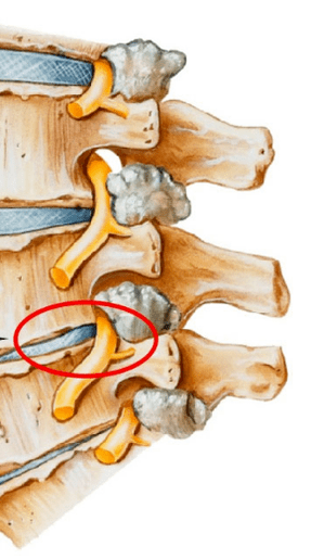 Diseases of the internal organs are the wrong position of the internal organs, their displacement and reduction due to various dysfunctions lead to a disruption of the overall equilibrium in the body. As a result, this is sharply affecting the position of the spine - the cervical, the lumbar vertebrae are displaced and deformed, leading to the corresponding species of osteochondrosis.
In general, osteochondrosis of the cervical region develops due to the effects of adverse external factors that disrupt the natural equilibrium position of the spine and other systems of the human body. Diagnostics
The diagnosis of cervical osteochondrosis begins with the collection of all the necessary information about the patient. The specialist asks for complaints that bother the person, is interested in his professional activities, as well as how he spends his weekend. An important point is the presence of osteochondrosis in parents, grandparents, because it is a hereditary disease.
The doctor then goes directly to the patient's visual examination. He studies the cervical ward and his back to the curvature of the stand, palpate the cervical region. This allows a specialist to evaluate the degree of development of the disease, since in advanced cases palpation of the cervical region causes acute pain.
When viewing, you should pay attention:on the severity of cervical lordosis;shoulder height in the patient;the possibility of asymmetry of the zones of the sickens;the possibility of asymmetry of the neck (such as a consequence of congenital pathology or acute muscle spasm);The condition of the muscles of the shoulder girdle and upper limbs (for example, single muscular atrophy may indicate compression of the cervical spine);The location of the chin - the chin is normal, must be located on the midline;The movement of the neck (burning bending, tilting to the right and rotation and rotation).
Palpation is carried out in the original position of the patient:lies on the back;lies on the stomach;Sitting in a chair.
The volume of movement volume is also carried out. It is performed in the original position of the patient sitting in a chair (to fix another spine).
Distribute the following major movements in the cervical region: flexion;extension;slopes to the right and left;Rotation.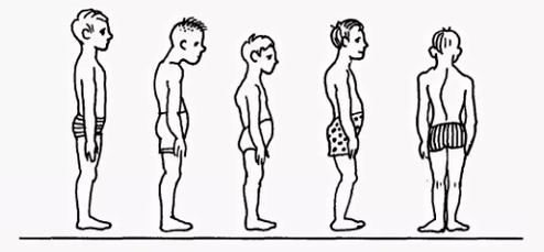
About half of the volume of flexion and lengthening occurs between the back of the head, the vertebrae of C1 and C2. The rest of the movement is carried out due to the main vertebrae, with a large scale of the movements in the vertebrae C5-C7. The lateral inclinations are distributed evenly between all vertebrae.
To make an accurate diagnosis, additional studies are prescribed:X -Ray of the cervical region. This method is suitable in the early stages of the disease, but may be useless in advanced forms.CT (computed tomography). It allows you to see structural changes in the vertebrae, but with the help of this method it is impossible to determine the size of the hernia between the vertebrae.MRI. It is considered to be the most effective method for diagnosing cervical osteochondrosis. You can determine the size of the hernia between the discs as well as the degree of their development.The doctor may also prescribe duplex scanning, which allows you to determine a violation of normal blood circulation in the arteries.Treatment
Treatment of cervical osteochondrosis is a complex therapy that involves taking medicines, gels, as well as various physiotherapy measures. An important role is played by therapeutic gymnastics as well as massage of the problem area.Medication
It is impossible to eliminate the consequences of degenerative-dystrophic changes in the spine without the use of medication. The use of medicines in the treatment of osteochondrosis of the cervical region is one of the main points of the complex approach to healing used by most specialists in traditional medicine.
It is intended to solve several problems, including:Stop the symptom of pain and eliminate the inflammatory process;Remove muscle spasm;stimulates the process of regeneration of cartilage and bone tissue cells;strengthening the protective properties of the body, increased immunity;Improve the general condition by eliminating other symptoms that prevent recovery.
Depending on these purposes, all medicines prescribed by a doctor suffering from cervical osteochondrosis may be conditionally divided into the following groups:Analgesics (non -steroidal drugs that relieve pain).Anti -inflammatory (steroid) are hormonal drugs that relieve inflammatory phenomena and thus eliminate pain.Chondroprotectors are medicines containing substances that replace the components of cartilage tissue - chondroitin, hyaluronic acid.Mussorelaxants. These are medicines that relax muscle tone. They are used in surgery and orthopedics as ancillary drugs to stop pain. Such medicines are administered by Parentl and therefore always under the supervision of a doctor. Vitamins. Osteochondrosis of the cervical region prescribes vitamins, has a beneficial effect on the peripheral nervous system and improves conductivity. Water -soluble vitamins: B1, B6, B12, fat soluble vitamins: A, C, E, E. In recent years, medicines containing both painkillers and vitamins components have been combined.Ointments and gels for outdoor use.Physiotherapy
The main purpose of physiotherapy treatment is to stimulate the process of regeneration in the body and to eliminate pain. The most popular methods in the treatment of cervical chondrosis are as follows:Ultrasound. Ultrasonic wave physicia is used to relieve serious pain and inflammatory reaction. Ultrasound is a massage of the tissue of the neck, after which the metabolism is activated. Vibration massage. The impact on the area of pain during the vibration massage is through mechanical oscillatory movements. For the proper behavior of this physiotherapy, a strip vibrator is usually used.Electrophoresis. This method of conducting physiotherapy care for cervical osteochondrosis is carried out using diadynamic and modulated currents, as well as electric fields when the body is administered in the tissues of medicines. Electrophoresis perfectly relieves spasmodic syndromes and eliminates pain in inflamed muscles.Magnetotherapy. The essence of magnetotherapy such as a physiosterization in osteochondrosis of the cervical region is explained by the use of permanent or variables of magnet fields, frequency of different sizes. This method can help the patient eliminate the pain and stop the inflammatory process in the hearth. The procedure is often performed at home after acquiring a special device with a magnetographer.Dutoenzor-Terapy. At present, a very popular method of physiotherapy, consisting in stretching the spine under the mass of the patient's body. In order to perform such a procedure, a mattress, arranged in a special way that has inclined ribs is required and they change the location below the weight of their own body. Muscle tone normalizes, leading to their relaxation.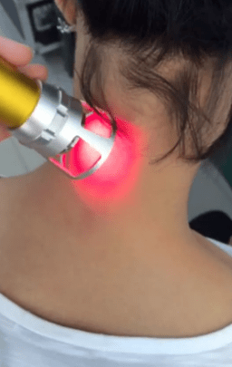 Laser therapy. The laser has a complex effect on the focus of inflammation, it activates biological processes in the tissues of the nervous system. This allows you to get a positive effect of treatment. The complex effect on the body consists in the effect of anti -inflammatory, analgesic and wound healing. A laser treatment procedure should not exceed 15 minutes. This is the optimal time of the Helic-Neon laser exposure to the affected areas. In this case, the duration of the laser on one pain should not exceed 2 minutes. Balneotherapy. The benefits of mineral water have long been known, this is based on balneotherapy. The procedure involves the active use of water resources in the treatment of osteochondrosis. In addition to taking baths, different types of souls and active swimming in the pool, therapy involves the use of therapeutic mud applications in painful areas of the body. The therapeutic effect is achieved by the simultaneous effect of chemically active substances contained in water under different temperature conditions. The technique allows you to stop the pain syndrome by improving local microcirculation in the tissues.Exercise therapy
It should be remembered that exercise therapy does not take place when signs of exacerbation begin: pain. After the LFK complex, they can be strengthened and cause inconvenience.
There are a number of general billing recommendations:Physical upbringing should be carried out indoors with good ventilation, an excellent street version.The classes are performed only during the remission of the disease (when there are no symptoms).Exercise therapy is thought to be wide, not disturbing movements and breathing.All movements are smooth, the amplitude and the number of repetitions gradually increase.If the pain starts, you should stop the lesson immediately.The classes and terminate pressure and pulse measurements are preceded. When these indicators differ from normal, the load should be reduced.It is advisable to listen to your breathing throughout the lesson, this will increase efficiency. All stretching exercises are performed when exhaled.It is very important to gradually increase the load and the number of repetitions, this will reduce the risk of injury and prevent fatigue. 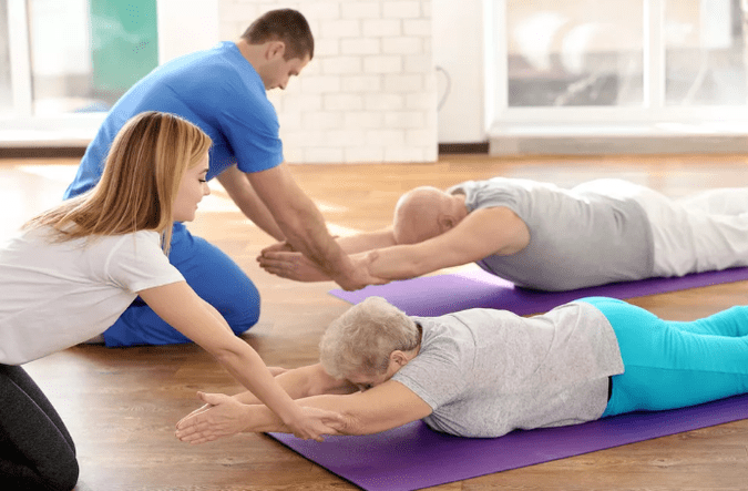 Exercise is important for regular performance, so you can achieve a quick result.Before you begin independent classes, you should consult a doctor and agree with him a set of exercises.
Recommended exercises in the starting position lying on the stomach:The head is ultimately on the forehead, with your hands on the back of the head, the elbows parallel to the floor. Lift your head with your hands off the floor, hold this position to 4 accounts, lower and relax. Repeat 2-4 times.The head is on the stop of the chin, the palms under the chin. Time in time, stretch your hands forward, two - spread on the side, three - stretch forward, four - the starting position. Repeat 2-4 times.Hands stretched forward. Swam swimming style, repeated 4-8 times.Palm under your chin, an accent on the palm of your forehead. Alternatively, remove the heel of the ass. Repeat 4-8 times.
Recommended exercises in the starting position lying on the side (right, then on the left):The right hand is extended, the right ear is on it, lift the right arm with your head, hold the position to 4 accounts, lower and relax. Repeat 2-4 times.The left hand rests on the floor in front of the chest, the left leg makes the movements of the fly back -behind. Repeat 6-8 times.The left hand on the body, lift your left hand up and out, a slower, a lower one. Repeat 2-4 times.The left hand of the thigh. By pulling the two knees to your chest when exhaled, straighten your feet inspiration. Repeat the exercises 2-4 times.


